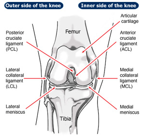
The Knee Joint The knee joint is a complex hinge joint that also allows some rotation as the knee bends or flexes. The knee joint is a complex hinge joint that also allows some rotation as the knee bends or flexes.
The upper part of the knee is formed by the distal (lowermost) end of the femur which in turn has two prominences; the medial (inner) and lateral (outer) femoral condyles. The lower part of the knee is formed from the proximal (upper) tibia and has similar medial and lateral tibial prominences or plateau. In the centre of the joint are two smaller prominences which are insertion points for the internal or cruciate ligaments of the knee. The anterior cruciate ligament (ACL) is closer to the front on the tibia and helps prevent forward movement of the tibia on the femur and the posterior cruciate ligament (PCL) lies further back and helps prevent the tibia riding backwards on the femur and also acts as a check rein to hyperextension. On sideways or lateral X-ray the patella (kneecap) can be seen. The patella is actually a bone within a tendon (a sesamoid bone), connecting to the tibia by the patella tendon. On its proximal (uppermost) surface the quadriceps muscle inserts. On the tangential or skyline view the patella is seen in profile and the 'V' shaped undersurface can be seen. There is a corresponding groove in the femur (the trochlear) for the patella to run up and down in as the knee flexes. As the knee moves forwards and backwards into flexion and extension there are also subtle rotational movements inside the knee.
|